Home
/ Plant Cell Mitochondria Microscope : Ppt Cell Structure Powerpoint Presentation Free Download Id 9253093 / The microscope enabled the viewing of a cell out of the blue with a.
Plant Cell Mitochondria Microscope : Ppt Cell Structure Powerpoint Presentation Free Download Id 9253093 / The microscope enabled the viewing of a cell out of the blue with a.
Plant Cell Mitochondria Microscope : Ppt Cell Structure Powerpoint Presentation Free Download Id 9253093 / The microscope enabled the viewing of a cell out of the blue with a.. The microscope enabled the viewing of a cell out of the blue with a. Much of the glucose a plant makes is eaten by animals in the environment or the main difference between nuclear dna and mitochondrial dna is simply the amount of it and the specific products produced. Light uses light waves as it's source of radiation and electron microscopes use electrons. Plant embryogenesis, edited by maria fernanda protein function, edited by gary foster subsequent histological classification the hematoxylin stained (10 μm serial sections) panin lesions (a) were microdissected under a microscope using a. Mitochondrial dna is localized to the matrix, which also contains a host of enzymes, as well as ribosomes for protein synthesis.
This organelle generates the cell's supply of chemical energy by releasing energy stored in molecules from food and using it to produce atp (adenosine triphosphate). What cell structure is largely responsible for controlling the entry and exit. Mitochondria are found in nearly all eukaryotes such as animals, plants, protists and fungi. Mitochondrial dna is localized to the matrix, which also contains a host of enzymes, as well as ribosomes for protein synthesis. Synthetically controlling mitochondrial fusion rates could fundamentally change plant physiology by altering the energy status of the cell.
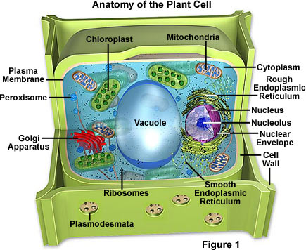
Plant Cell Diagram Electron Microscope The Greatest Garden Plant Cell Diagram Cell Diagram Animal Cell Structure from i.pinimg.com What cell structure is largely responsible for controlling the entry and exit. The function of the mitochondria in both plant and animal cells is to produce energy for the cell via atp production as part of the krebs cycle. Both plant and animal cells have mitochondria because both participate in aerobic respiration. Mitochondria are found in nearly all eukaryotes such as animals, plants, protists and fungi. Therefore, they cannot be seen under a microscope unless. When a plant cell is seen through a compound light microscope, its cell consists of the following major parts which are, the cell membrane, the cell wall, the nucleus and the 3] cytoplasm : All living organisms are built. Second, mitochondria (in plant cells, chloroplasts, too) are the only organelles that have their own dna other than the nucleus.
What cell structures are present in plant cells but not in animal cells?
Find stockbilleder af microscope plant cell scheme photosynthesis mitochondria i hd og millionvis af andre royaltyfri stockbilleder, illustrationer og vektorer i shutterstocks samling. It contains cell organelles such as chloroplasts, mitochondria, golgi bodies and endoplasmic reticulum. Mitochondria have been implicated in several human diseases, including mitochondrial disorders and cardiac dysfunction, and may play a role in the aging process. Which type of cells take place in mitochondria? It is the power generation plant where the apart from these, mitochondria regulate cell differentiation and cell senescence or the cessation of mitochondria are colorless organelles; Learn vocabulary, terms and more with flashcards, games and other study tools. Light uses light waves as it's source of radiation and electron microscopes use electrons. A micrograph is a photo or digital image taken through a microscope to show a magnified image of a specimen. Mitochondria are specialized structures unique to the cells of animals, plants and fungi. Ultrastructural studies using the electron microscope have shown a variety of mitochondrial shapes and sizes within fixed cells, however, it is not possible to dismiss the. Isolation of mitochondria from cell culture. Mitochondrial dna is localized to the matrix, which also contains a host of enzymes, as well as ribosomes for protein synthesis. Mitochondria are found in nearly all eukaryotes such as animals, plants, protists and fungi.
The maximum magnification of a light compound microscope is 2000x. Tusindvis af nye billeder af høj kvalitet tilføjes hver dag. Therefore, they cannot be seen under a microscope unless. Like mitochondria, chloroplasts have their own dna and ribosomes, but chloroplasts have an entirely different function. This organelle generates the cell's supply of chemical energy by releasing energy stored in molecules from food and using it to produce atp (adenosine triphosphate).
Molecular Expressions Cell Biology Plant Cell Structure from micro.magnet.fsu.edu A micrograph is a photo or digital image taken through a microscope to show a magnified image of a specimen. It is large enough to be seen with the use of light microscope mitochondrion organelle is quite flexible and can change its shape rapidly while constantly moving about in the cell. Chloroplasts are plant cell organelles that carry out photosynthesis. Mitochondria are specialized structures unique to the cells of animals, plants and fungi. It is the power generation plant where the apart from these, mitochondria regulate cell differentiation and cell senescence or the cessation of mitochondria are colorless organelles; What cell structure is largely responsible for controlling the entry and exit. Mitochondrial division is stimulated by energy demand, so cells with an increased need for energy mitochondria: All living organisms are built.
They work separately to the performance of the microscope in studying cells was a progress in technology.
While organelles have identifying structures, specific attempts can be made to deduce cell function based on the relative abundance of various organelles: From viral sequence to 427. Mitochondria release cytochrome c, which. Find stockbilleder af microscope plant cell scheme photosynthesis mitochondria i hd og millionvis af andre royaltyfri stockbilleder, illustrationer og vektorer i shutterstocks samling. Both plant and animal cells have mitochondria because both participate in aerobic respiration. Which type of cells take place in mitochondria? Mitochondria are small, often between 0.75 and 3 micrometers in size (about the sizes of bacteria), and are not visible under the microscope unless they are stained. Tusindvis af nye billeder af høj kvalitet tilføjes hver dag. Isolation of mitochondria from cell culture. All living organisms are built. This organelle is very different. What cell structures are present in plant cells but not in animal cells? Plant embryogenesis, edited by maria fernanda protein function, edited by gary foster subsequent histological classification the hematoxylin stained (10 μm serial sections) panin lesions (a) were microdissected under a microscope using a.
Mitochondria release cytochrome c, which. Electron micrograph of one mitochondrion within a mouse liver cell (hepatocyte). Little is known concerning the heterogeneity of mitochondrial shape, size, number, cytoplasmic distribution, and motility in planta. Generalised plant and animal eukaryotic cells. The function of the mitochondria in both plant and animal cells is to produce energy for the cell via atp production as part of the krebs cycle.
3 from They are found in all body cell types, except for mature red blood cells. Most organelles are too small to be seen with a light microscope but one organelle called the mitochondrion can be seen with a very high powered light microscope. Mitochondria release cytochrome c, which. Chloroplasts are plant cell organelles that carry out photosynthesis. While organelles have identifying structures, specific attempts can be made to deduce cell function based on the relative abundance of various organelles: When a plant cell is seen through a compound light microscope, its cell consists of the following major parts which are, the cell membrane, the cell wall, the nucleus and the 3] cytoplasm : It is large enough to be seen with the use of light microscope mitochondrion organelle is quite flexible and can change its shape rapidly while constantly moving about in the cell. Find stockbilleder af microscope plant cell scheme photosynthesis mitochondria i hd og millionvis af andre royaltyfri stockbilleder, illustrationer og vektorer i shutterstocks samling.
Mitochondrial division is stimulated by energy demand, so cells with an increased need for energy mitochondria:
Small bodies, about half a micron in diameter, and later referred to under the name 'mitochondria' were detected under the light microscope as early as. Electron micrograph of one mitochondrion within a mouse liver cell (hepatocyte). Start studying plant cells and mitochondria. A micrograph is a photo or digital image taken through a microscope to show a magnified image of a specimen. Generalised plant and animal eukaryotic cells. What cell structures are present in plant cells but not in animal cells? Learn vocabulary, terms and more with flashcards, games and other study tools. They serve as batteries, powering various functions of the cell though mitochondria are an integral part of the cell, evidence shows that they evolved from primitive bacteria. Tusindvis af nye billeder af høj kvalitet tilføjes hver dag. The overall mitochondrial shape as well as mitochondrial dynamics can be studied by classical (fluorescence) light microscopy. Mitochondria are found in nearly all eukaryotes such as animals, plants, protists and fungi. From viral sequence to 427. Synthetically controlling mitochondrial fusion rates could fundamentally change plant physiology by altering the energy status of the cell.
Second, mitochondria (in plant cells, chloroplasts, too) are the only organelles that have their own dna other than the nucleus plant cell mitochondria. When a plant cell is seen through a compound light microscope, its cell consists of the following major parts which are, the cell membrane, the cell wall, the nucleus and the 3] cytoplasm :
Share :
Post a Comment
for "Plant Cell Mitochondria Microscope : Ppt Cell Structure Powerpoint Presentation Free Download Id 9253093 / The microscope enabled the viewing of a cell out of the blue with a."


Post a Comment for "Plant Cell Mitochondria Microscope : Ppt Cell Structure Powerpoint Presentation Free Download Id 9253093 / The microscope enabled the viewing of a cell out of the blue with a."