Home
/ Animal Cell Under Electron Microscope Labelled - Topic 1.2 Ultra-Structure of Cells - AMAZING WORLD OF ... : Smooth endoplasmic reticulum, mitochondria, golgi bodies, lysosomes.
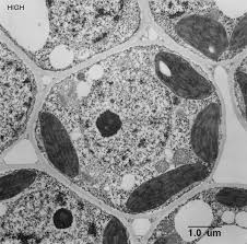
Animal Cell Under Electron Microscope Labelled - Topic 1.2 Ultra-Structure of Cells - AMAZING WORLD OF ... : Smooth endoplasmic reticulum, mitochondria, golgi bodies, lysosomes.
Animal Cell Under Electron Microscope Labelled - Topic 1.2 Ultra-Structure of Cells - AMAZING WORLD OF ... : Smooth endoplasmic reticulum, mitochondria, golgi bodies, lysosomes.. Bring your presentation to life. The diagram is very clear, and labeled the diagram is very clear, and labeled; Image:animal cell seen under electron microscope. Students will observe onion cells under a these cells have a dark stained nucleus and a large vacuole in the centre. All information about labeled plant cell under electron microscope.
A cell is a very tiny structure which exists in living bodies. Illustrated in figure 2 are a pair of fibroblast deer skin cells that have been labeled with fluorescent probes and photographed in the microscope to reveal. This site is using cookies under cookie policy. Can distinguish between objects as small as 200nm. The detail that can be seen, or resolution, is also important.
labeled animal cell under electron microscope 8745961 orig ... from labels-top.com But at the same time it is interpretive. Light and electron microscopes allow us to see inside cells. Image:animal cell seen under electron microscope. Under a high power microscope like the scanning transmission electron microscope, it is possible even to stain and observe the detailed. Labeled animal cell under electron illustrate only a plant cell as seen under electron. Ishita observed a slide of eukaryotic cell under electron microscope. All information about labeled plant cell under electron microscope. Now the first thing to point out when looking at images under an electron microscope.
After completing this section, you should know:
As for seeing electrons under any microscope in general, i would say we have come as close to it as scientifically and technically possible with the tem having a resolution of 2 nm (there might be more advanced tools with here is an electron micrograph of an animal cell with the labels superimposed The role and function of the plasma membrane; Under the electron microscope, other parts of the cell can be seen, each performing a specific function. Image:animal cell seen under electron microscope. This site is using cookies under cookie policy. However, the internal structure and organelles are more or less similar. All information about labeled plant cell under electron microscope. But at the same time it is interpretive. It also has a very high resolving power. Resolving power is the ability to distinguish between separate things which are close to each other. During the last 70 years, transmission electron microscopy (tem) has developed our knowledge about ultrastructure of the cells and tissues. Now the first thing to point out when looking at images under an electron microscope is the scale. Penguins global warming, dove soap bar, bugatti veyron super sport.
As for seeing electrons under any microscope in general, i would say we have come as close to it as scientifically and technically possible with the tem having a resolution of 2 nm (there might be more advanced tools with here is an electron micrograph of an animal cell with the labels superimposed 25.02.2021 · leaf cell under microscope labeled written by macpride monday, april 13, 2020 add comment edit. Cytoplasm, ribosomes, rough endoplasmic reticulum; Image:animal cell seen under electron microscope. Labels are a means of identifying a product or container through a piece of fabric, paper, metal or plastic film onto which information about them is printed.
Topic 1.2 Ultra-Structure of Cells - AMAZING WORLD OF ... from www.mrgscience.com Animal and plant cell under electron microscope. At the end of the activity, the student should be able to: Animal cell electron micrograph labelling. Electron microscopes use electron beams focused by electromagnets to magnify and resolve microscopic specimens. Its leaves are only two cells thick, making it possible to easily. Smooth endoplasmic reticulum, mitochondria, golgi bodies, lysosomes. Light and electron microscopes allow us to see inside cells. As the wavelength of an electron can be up to 100.
Under a high power microscope like the scanning transmission electron microscope, it is possible even to stain and observe the detailed.
Animal cell electron micrograph labelling. Exocrine cell of pancreas electron micro… Draw a labelled diagram of an onion epidermal cell seen under the microscope. Penguins global warming, dove soap bar, bugatti veyron super sport. Typical animal cell pinocytotic vesicle lysosome golgi vesicles golgi vesicles rough er (endoplasmic reticulum) smooth er (no ribosomes) cell (plasma) membrane… 2. Under the microscope, animal cells appear different based on the type of the cell. Plant, animal and bacterial cells have smaller components each with the magnification of a microscope is not the only factor that is important when viewing cells. Identify the basic structures of a cell 2. After this, add another oval shape outside the line you just drew, and this will make the cell membrane to your animal cell. Image:plant cell seen under electron microscope. Its leaves are only two cells thick, making it possible to easily. That cells can be of different shapes and sizes. See how a generalized structure of an animal cell and plant cell look with labeled diagrams.
All information about labeled plant cell under electron microscope. Its leaves are only two cells thick, making it possible to easily. Exocrine cell of pancreas electron micro… The ability to visualise columns of atoms under a transmission electron microscope indicates how extremely powerful and high resolution these instruments are. Electron microscopes use electron beams focused by electromagnets to magnify and resolve microscopic specimens.
labeled animal cell under electron microscope 8745961 orig ... from labels-top.com All information about labeled plant cell under electron microscope. 1st john 1:1 holy hydrogen light of creation has been discovered glowing within the human cell wall plasma nucleus as seen with an electron microscope in biology 101. Identify the basic structures of a cell 2. Here's a diagram of a plant cell: Image:animal cell seen under electron microscope. Animal cell electron micrograph labelling. Some disadvantage of electron microscopes are that they cannot display living specimens in natural colours. Ishita observed a slide of eukaryotic cell under electron microscope.
Ishita observed a slide of eukaryotic cell under electron microscope.
Under a high power microscope like the scanning transmission electron microscope, it is possible even to stain and observe the detailed. A cell is a very tiny structure which exists in living bodies. Now the first thing to point out when looking at images under an electron microscope. Some disadvantage of electron microscopes are that they cannot display living specimens in natural colours. Typical animal cell pinocytotic vesicle lysosome golgi vesicles golgi vesicles rough er (endoplasmic reticulum) smooth er (no ribosomes) cell (plasma) membrane… 2. Resolving power is the ability to distinguish between separate things which are close to each other. Image:animal cell seen under electron microscope. In truth, there are still features of plant and animal cells we're only lately. Identify the different cell shape and relate it to the cell's function. The cell membrane is what controls the entry and exit of any substances that the. Students will observe onion cells under a these cells have a dark stained nucleus and a large vacuole in the centre. 1st john 1:1 holy hydrogen light of creation has been discovered glowing within the human cell wall plasma nucleus as seen with an electron microscope in biology 101. As the wavelength of an electron can be up to 100.
Share :
Post a Comment
for "Animal Cell Under Electron Microscope Labelled - Topic 1.2 Ultra-Structure of Cells - AMAZING WORLD OF ... : Smooth endoplasmic reticulum, mitochondria, golgi bodies, lysosomes."


Post a Comment for "Animal Cell Under Electron Microscope Labelled - Topic 1.2 Ultra-Structure of Cells - AMAZING WORLD OF ... : Smooth endoplasmic reticulum, mitochondria, golgi bodies, lysosomes."